
What is a tongue-tie? What parents need to know

The tongue is secured to the front of the mouth partly by a band of tissue called the lingual frenulum. If the frenulum is short, it can restrict the movement of the tongue. This is commonly called a tongue-tie.
Children with a tongue-tie can’t stick their tongue out past their lower lip, or touch their tongue to the top of their upper teeth when their mouth is open. When they stick out their tongue, it looks notched or heart-shaped. Since babies don’t routinely stick out their tongues, a baby’s tongue may be tied if you can’t get a finger underneath the tongue.
How common are tongue-ties?
Tongue-ties are common. It’s hard to say exactly how common, as people define this condition differently. About 8% of babies under age one may have at least a mild tongue-tie.
Is it a problem if the tongue is tied?
This is really important: tongue-ties are not necessarily a problem. Many babies, children, and adults have tongue-ties that cause them no difficulties whatsoever.
There are two main ways that tongue-ties can cause problems:
- They can cause problems with breastfeeding by making it hard for some babies to latch on well to the mother’s nipple. This causes difficulty with feeding for the baby and sore nipples for the mother. It doesn’t happen to all babies with a tongue-tie; many of them can breastfeed successfully. Tongue-ties are not to blame for gassiness or fussiness in a breastfed baby who is gaining weight well. Babies with tongue-ties do not have problems with bottle-feeding.
- They can cause problems with speech. Some children with tongue-ties may have difficulty pronouncing certain sounds, such as t, d, z, s, th, n, and l. Tongue-ties do not cause speech delay.
What should you do if think your baby or child has a tongue-tie?
If you think that your newborn is not latching well because of a tongue-tie, talk to your doctor. There are many, many reasons why a baby might not latch onto the breast well. Your doctor should take a careful history of what has been going on, and do a careful examination of your baby to better understand the situation.
You should also have a visit with a lactation specialist to get help with breastfeeding — both because there are lots of reasons why babies have trouble with latching on, and also because many babies with a tongue-tie can nurse successfully with the right techniques and support.
Talk to your doctor if you think that a tongue-tie could be causing problems with how your child pronounces words. Many children just take some time to learn to pronounce certain sounds. It is also a good idea to have an evaluation by a speech therapist before concluding that a tongue-tie is the problem.
What can be done about a tongue-tie?
When necessary, a doctor can release a tongue-tie using a procedure called a frenotomy. A frenotomy can be done by simply snipping the frenulum, or it can be done with a laser.
However, nothing should be done about a tongue-tie that isn’t causing problems. While a frenotomy is a relatively minor procedure, complications such as bleeding, infection, or feeding difficulty sometimes occur. So it’s never a good idea to do it just to prevent problems in the future. The procedure should only be considered if the tongue-tie is clearly causing trouble.
It’s also important to know that clipping a tongue-tie doesn’t always solve the problem, especially with breastfeeding. Studies do not show a clear benefit for all babies or mothers. That’s why it’s important to work with a lactation expert before even considering a frenotomy.
If a newborn with a tongue-tie isn’t latching well despite strong support from a lactation expert, then a frenotomy should be considered, especially if the baby is not gaining weight. If it is done, it should be done early on and by someone with training and experience in the procedure.
What else should parents know about tongue-tie procedures?
Despite the fact that the evidence for the benefits of frenotomy is murky, many providers are quick to recommend them. If one is being recommended for your child, ask questions:
- Make sure you know exactly why it is being recommended.
- Ask whether there are any other options, including waiting.
- Talk to other health care providers on your child’s care team, or get a second opinion.
About the Author

Claire McCarthy, MD, Senior Faculty Editor, Harvard Health Publishing
Claire McCarthy, MD, is a primary care pediatrician at Boston Children’s Hospital, and an assistant professor of pediatrics at Harvard Medical School. In addition to being a senior faculty editor for Harvard Health Publishing, Dr. McCarthy … See Full Bio View all posts by Claire McCarthy, MD
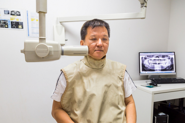
Ready to give up the lead vest?

At a dental appointment last month, I spotted a lead vest hanging unassumingly on the wall of the exam room as soon as I walked in. “Still there, but now obsolete,” I thought.
I’d just learned about new guidelines from the American Dental Association (ADA) saying lead vests and thyroid collars that cover the neck are no longer needed during dental x-rays. But they’d been a fixture of my dental experiences — including many cavities, four root canals, a tooth extraction, and two crowns — for my entire life. What changed, and could I feel safe without the vest?
Why were lead vests used in past years?
Lead vests and thyroid collars have been worn by countless Americans during dental x-rays over the years. They’ve been in use for far longer than my lifetime — about 100 years. The heavy apron-like shields are placed over sensitive areas, including the chest and neck, before the x-rays are taken.
“I haven’t worn a lead apron in the last 10 or 15 years — unless a dentist insists I put it on — because I know it isn’t needed,” says Dr. Bernard Friedland, an associate professor of oral medicine, infection, and immunity at Harvard School of Dental Medicine.
What has changed about dental x-rays?
When lead vests and thyroid collars were first recommended, x-ray technology was much less precise. But the technology has evolved significantly over the last few decades in ways that dramatically improve patient safety:
- Digital x-rays enable far smaller radiation doses, reducing radiation exposure and the risks associated with higher doses, such as cancer. “The doses used in dental radiology are negligibly small now. If you go to the dentist today for a full series of mouth x-rays that are taken with a digital sensor, the total exposure time is just over five seconds,” explains Dr. Friedland, an expert in oral radiology. “A hundred or so years ago, that exposure time would have been many minutes.”
- The small size of today’s x-ray beam significantly reduces radiation “scatter” and restricts the beam size to only the area needing to be imaged. This protects patients from radiation exposure to other parts of the body.
A less-recognized strike against using lead vests and thyroid collars is their ability to get in the way. They may block the primary x-ray beam, preventing dentists from capturing needed images. This quirk can lead to repeat imaging and unnecessary exposure to additional radiation. This is more likely to occur with panoramic x-rays.
The gear may also spread germs, Dr. Friedland notes. Although disinfected, it’s not sterilized between uses. “There’s a risk of spreading bacteria and viruses,” he says. “To me, that’s also an issue and another reason I don’t want to use one on myself.”
Who no longer needs the shields?
No one does — even children, who presumably have a long life of dental x-rays in front of them. The new recommendations apply to all patients regardless of age, health status, or pregnancy, the ADA says.
The recommendation to discontinue lead vests has been a long time in the making. In fact, the ADA isn’t the first professional organization to propose it. The American Association of Physicists in Medicine did so in 2019, followed by the American College of Radiology in 2021 and the American Academy of Oral and Maxillofacial Radiology in 2023.
Are some people confused or concerned about the no-lead-vest policy?
Yes. The new guidelines are bound to draw confusion and fear, Dr. Friedland says. Some people may even insist on continuing to wear a lead vest during x-rays.
“A big problem is that people’s perception of risk is very skewed,” he says. “Some people, you’ll never convince.”
People are likely to feel more comfortable if the practice is uniformly adopted by dentists. However, the ability to implement this change may hinge partly on public response. And it could take a while to fully adopt.
“I think the public is going to have more say on this than dentists,” Dr. Friedland says. “It might take a generation to make this change, maybe longer.”
Still concerned about the new recommendations?
If you have lingering concerns about the new recommendations, talk to your dentist.
And ask if dental x-rays are necessary to proceed with your diagnosis or treatment plan. Sometimes it’s possible to take fewer x-rays — such as bitewing x-rays of the upper and lower back teeth only — or to use certain types of imaging less frequently. Even with far safer x-ray conditions, dentists should be able to justify that the information from images is integral to diagnose problems or improve care, Dr. Friedland says.
It’s worth noting that the dose of radiation, while far lower than in the past, varies with the type of imaging and which parts of the jaw are being imaged. For example, the digital dental x-rays mentioned above involve less radiation than conventional dental x-rays. Either panoramic dental x-rays, or 3-D dental x-rays taken with a CBCT system that rotates around the head, typically involve more radiation than conventional dental x-rays.
Whenever possible, dentists should use images taken during previous dental exams, according to the ADA. “If I don’t need an x-ray, I don’t get one,” says Dr. Friedland. “I’m not cavalier about it. I also use technical parameters that keep the x-ray dose as low as reasonably possible.”
About the Author

Maureen Salamon, Executive Editor, Harvard Women's Health Watch
Maureen Salamon is executive editor of Harvard Women’s Health Watch. She began her career as a newspaper reporter and later covered health and medicine for a wide variety of websites, magazines, and hospitals. Her work has … See Full Bio View all posts by Maureen Salamon
About the Reviewer
Howard E. LeWine, MD, Chief Medical Editor, Harvard Health Publishing
Dr. Howard LeWine is a practicing internist at Brigham and Women’s Hospital in Boston, Chief Medical Editor at Harvard Health Publishing, and editor in chief of Harvard Men’s Health Watch. See Full Bio View all posts by Howard E. LeWine, MD
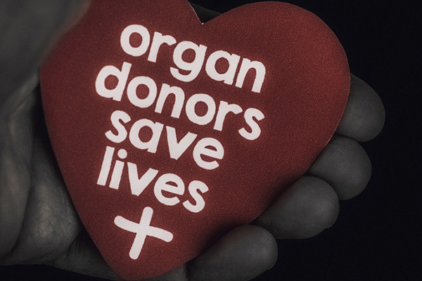
Flowers, chocolates, organ donation — are you in?

Chocolates and flowers are great gifts for Valentine’s Day. But what if the gifts we give then or throughout the year could be truly life-changing? A gift that could save a life or free someone from dialysis?
You can do this. For people in need of an organ, tissue, or blood donation, a donor can give them a gift that exceeds the value of anything that you can buy. Fittingly, Valentine’s Day is also known as National Donor Day, a time for blood drives and sign-ups for organ and tissue donation. Have you ever wondered what can be donated? Had reservations about donating after death or concerns about risks for live donors? Read on.
The enormous impact of organ, tissue, or cell donation
Imagine you have kidney failure requiring dialysis 12 or more hours each week just to stay alive. Even with this, you know you’re still likely to die a premature death. Or, if your liver is failing, you may experience severe nausea, itching, and confusion; death may only be a matter of weeks or months away. For those with cancer in need of a bone marrow transplant, or someone who’s lost their vision due to corneal disease, finding a donor may be their only good option.
Organ or tissue donation can turn these problems around, giving recipients a chance at a long life, a better quality of life, or both. And yet, the number of people who need organ donation far exceeds compatible donors. While national surveys have found about 90% of Americans support organ donation, only 40% have signed up. More than 103,000 women, men, and children are awaiting an organ transplant in the US. About 6,200 die each year, still waiting.
What can you donate?
The list of ways to help has grown dramatically. Some organs, tissues, or cells can be donated while you’re alive; other donations are only possible after death. A single donor can help more than 80 people!
After death, people can donate:
- bone, cartilage, and tendons
- corneas
- face and hands (though uncommon, they are among the newest additions to this list)
- kidneys
- liver
- lungs
- heart and heart valves
- stomach and intestine
- nerves
- pancreas
- skin
- arteries and veins.
Live donations may include:
- birth tissue, such as the placenta, umbilical cord, and amniotic fluid, which can be used to help heal skin wounds or ulcers and prevent infection
- blood cells, serum, or bone marrow
- a kidney
- part of a lung
- part of the intestine, liver, or pancreas.
To learn more about different types of organ donations, visit Donate Life America.
Becoming a donor after death: Questions and misconceptions
Common misconceptions about becoming an organ donor limit the number of people who are willing to sign up. For example, many people mistakenly believe that
- doctors won’t work as hard to save your life if you’re known to be an organ donor — or worse, doctors will harvest organs before death
- their religion forbids organ donation
- you cannot have an open-casket funeral if you donate your organs.
None of these is true, and none should discourage you from becoming an organ donor. Legitimate medical professionals always keep the patient’s interests front and center. Care would never be jeopardized due to a person’s choices around organ donation. Most major religions allow and support organ donation. If organ donation occurs after death, the clothed body will show no outward signs of organ donation, so an open-casket funeral is an option for organ donors.
Live donors: Blood, bone marrow, and organs
Have you ever donated blood? Congratulations, you’re a live donor! The risk for live donors varies depending on the intended donation, such as:
- Blood, platelets, or plasma: If you’re donating blood or blood products, there is little or no risk involved.
- Bone marrow: Donating bone marrow requires a minor surgical procedure. If general anesthesia is used, there is a chance of a reaction to the anesthesia. Bone marrow is removed through needles inserted into the back of the pelvis bones on each side. Back or hip pain is common, but can be controlled with pain relievers. The body quickly replaces the bone marrow removed, so no long-term problems are expected.
- Stem cells: Stem cells are found in bone marrow or umbilical cord blood. They also appear in small numbers in our blood and can be donated through a process similar to blood donation. This takes about seven or eight hours. Filgrastim, a medication that increases stem cell production, is given for a number of days beforehand. It can cause side effects such as flulike symptoms, bone pain, and fatigue, but these tend to resolve soon after the procedure.
- Kidney, lung, or liver: Surgery to donate a kidney or a portion of a lung or liver comes with a risk of complications, reactions to anesthesia, and significant recovery time. It’s no small matter to give a kidney, or part of a lung or liver.
The vast number of live organ donations occur without complications, and donors typically feel quite positive about the experience.
Who can donate?
Almost anyone can donate blood cells –– including stem cells –– or be a bone marrow, tissue, or organ donor. Exceptions include anyone with active cancer, widespread infection, or organs that aren’t healthy.
What about age? By itself, your age does not disqualify you from organ donation. In 2023, two out of five people donating organs were over 50. People in their 90s have donated organs upon their deaths and saved the lives of others.
However, bone marrow transplants may fail more often when the donor is older, so bone marrow donations by people over age 55 or 60 are usually avoided.
Finding a good match: Immune compatibility
For many transplants, the best results occur when there is immune compatibility between the donor and recipient. Compatibility is based largely on HLA typing, which analyzes genetically-determined proteins on the surface of most cells. These proteins help the immune system identify which cells qualify as foreign or self. Foreign cells trigger an immune attack; cells identified as self should not.
HLA typing can be done by a blood test or cheek swab. Close relatives tend to have the best HLA matches, but complete strangers may be a good match as well.
Fewer donors among people with certain HLA types make finding a match more challenging. Already existing health disparities, such as higher rates of kidney disease among Black Americans and communities of color, are worsened by lower numbers of donors from these communities, an inequity partly driven by a lack of trust in the medical system.
The bottom line
You can make an enormous impact by becoming a donor during your life or after death. In the US, you must opt in to be a donor after death. (Research suggests the opt-out approach many other countries use could significantly increase rates of organ donation in this country.)
I’m hopeful that organ donation in the US and throughout the world will increase over time. While you can still go with chocolates for Valentine’s Day, maybe this year you can also go bigger and become a donor.
About the Author

Robert H. Shmerling, MD, Senior Faculty Editor, Harvard Health Publishing; Editorial Advisory Board Member, Harvard Health Publishing
Dr. Robert H. Shmerling is the former clinical chief of the division of rheumatology at Beth Israel Deaconess Medical Center (BIDMC), and is a current member of the corresponding faculty in medicine at Harvard Medical School. … See Full Bio View all posts by Robert H. Shmerling, MD

Discrimination at work is linked to high blood pressure

Experiencing discrimination in the workplace — where many adults spend one-third of their time, on average — may be harmful to your heart health.
A 2023 study in the Journal of the American Heart Association found that people who reported high levels of discrimination on the job were more likely to develop high blood pressure than those who reported low levels of workplace discrimination.
Workplace discrimination refers to unfair conditions or unpleasant treatment because of personal characteristics — particularly race, sex, or age.
How can discrimination affect our health?
“The daily hassles and indignities people experience from discrimination are a specific type of stress that is not always included in traditional measures of stress and adversity,” says sociologist David R. Williams, professor of public health at the Harvard T.H. Chan School of Public Health.
Yet multiple studies have documented that experiencing discrimination increases risk for developing a broad range of factors linked to heart disease. Along with high blood pressure, this can also include chronic low-grade inflammation, obesity, and type 2 diabetes.
More than 25 years ago, Williams created the Everyday Discrimination Scale. This is the most widely used measure of discrimination’s effects on health.
Who participated in the study of workplace discrimination?
The study followed a nationwide sample of 1,246 adults across a broad range of occupations and education levels, with roughly equal numbers of men and women.
Most were middle-aged, white, and married. They were mostly nonsmokers, drank low to moderate amounts of alcohol, and did moderate to high levels of exercise. None had high blood pressure at the baseline measurements.
How was discrimination measured and what did the study find?
The study is the first to show that discrimination in the workplace can raise blood pressure.
To measure discrimination levels, researchers used a test that included these six questions:
- How often do you think you are unfairly given tasks that no one else wanted to do?
- How often are you watched more closely than other workers?
- How often does your supervisor or boss use ethnic, racial, or sexual slurs or jokes?
- How often do your coworkers use ethnic, racial, or sexual slurs or jokes?
- How often do you feel that you are ignored or not taken seriously by your boss?
- How often has a coworker with less experience and qualifications gotten promoted before you?
Based on the responses, researchers calculated discrimination scores and divided participants into groups with low, intermediate, and high scores.
- After a follow-up of roughly eight years, about 26% of all participants reported developing high blood pressure.
- Compared to people who scored low on workplace discrimination at the start of the study, those with intermediate or high scores were 22% and 54% more likely, respectively, to report high blood pressure during the follow-up.
How could discrimination affect blood pressure?
Discrimination can cause emotional stress, which activates the body’s fight-or-flight response. The resulting surge of hormones makes the heart beat faster and blood vessels narrow, which causes blood pressure to rise temporarily. But if the stress response is triggered repeatedly, blood pressure may remain consistently high.
Discrimination may arise from unfair treatment based on a range of factors, including race, gender, religious affiliation, or sexual orientation. The specific attribution doesn’t seem to matter, says Williams. “Broadly speaking, the effects of discrimination on health are similar, regardless of the attribution,” he says, noting that the Everyday Discrimination Scale was specifically designed to capture a range of different forms of discrimination.
What are the limitations of this study?
One limitation of this recent study is that only 6% of the participants were nonwhite, and these individuals were less likely to take part in the follow-up session of the study. As a result, the study may not have fully or accurately captured workplace discrimination among people from different racial groups. In addition, blood pressure was self-reported, which may be less reliable than measurements directly documented by medical professionals.
What may limit the health impact of workplace discrimination?
At the organizational level, no studies have directly addressed this issue. Preliminary evidence suggests that improving working conditions, such as decreasing job demands and increasing job control, may help lower blood pressure, according to the study authors. In addition, the American Heart Association recently released a report, Driving Health Equity in the Workplace, that aims to address drivers of health inequities in the workplace.
Encouraging greater awareness of implicit bias may be one way to help reduce discrimination in the workplace. Implicit bias refers to the unconscious assumptions and prejudgments people have about groups of people that may underlie some discriminatory behaviors. You can explore implicit biases with these tests, which were developed at Harvard and other universities.
On an individual level, stress management training can reduce blood pressure. A range of stress-relieving strategies may offer similar benefits. Regularly practicing relaxation techniques or brief mindfulness reflections, learning ways to cope with negative thoughts, and getting sufficient exercise can help.
About the Author

Julie Corliss, Executive Editor, Harvard Heart Letter
Julie Corliss is the executive editor of the Harvard Heart Letter. Before working at Harvard, she was a medical writer and editor at HealthNews, a consumer newsletter affiliated with The New England Journal of Medicine. She … See Full Bio View all posts by Julie Corliss
About the Reviewer
Howard E. LeWine, MD, Chief Medical Editor, Harvard Health Publishing
Dr. Howard LeWine is a practicing internist at Brigham and Women’s Hospital in Boston, Chief Medical Editor at Harvard Health Publishing, and editor in chief of Harvard Men’s Health Watch. See Full Bio View all posts by Howard E. LeWine, MD

What complications can occur after prostate cancer surgery?

Earlier this year, US defense secretary Lloyd Austin was hospitalized for complications resulting from prostate cancer surgery. Details of his procedure, which was performed on December 22, were not fully disclosed. Press statements from the Pentagon indicated that Austin had undergone a minimally invasive prostatectomy, which is an operation to remove the prostate gland. Minimally invasive procedures are performed using robotic instruments passed through small “keyhole” incisions in the patient’s abdomen.
Just over a week later, Austin developed severe abdominal, hip, and leg pain. He was admitted to the intensive care unit at Walter Reed Hospital on January 2 for monitoring and further treatment. Doctors discovered that Austin had a urinary tract infection and fluid pooling in his abdomen that were impairing bowel functioning. The defense secretary was successfully treated, but then readmitted to the ICU on February 11 for what the Pentagon described as “an emergent bladder issue.” Two days after undergoing what was only described as a “non-surgical procedure performed under general anesthesia,” Austin was back at work. His cancer prognosis is said to be excellent.
Austin’s ordeal was covered extensively in the media. Although we cannot speculate about his specific case, to help our readers better understand the complications that might occur after a prostatectomy, I spoke with Dr. Boris Gershman, a urologist at Harvard-affiliated Beth Israel Deaconess Medical Center in Boston. Dr. Gershman is also a member of the advisory and editorial board for the Harvard Medical School Guide to Prostate Diseases.
How common are urinary tract infections after a prostatectomy?
Minimally invasive prostatectomy is generally well tolerated. In one study that examined complications among over 29,000 men who had the operation, the rate of urinary tract infections was only 2.1%. The risk of sepsis — a more serious condition that occurs if the body’s response to an infection damages other organs — is much lower than that.
How would a urinary tract infection occur?
Although urinary tract infections are rare after prostatectomy, bacteria can travel into the urinary system through a catheter. An important part of a prostatectomy involves connecting the urethra — which is a tube that carries urine out of the body — directly to the bladder after the prostate has been taken out. As a last step in that process, we pass a catheter [a soft silicone tube] through the urethra and into the bladder to promote healing. Infection risks are minimized by giving antibiotics both during surgery and then again just prior to removing the catheter one to two weeks after the operation.
How do you treat urinary infectious complications when they do happen?
It’s not unusual to find small amounts of bacteria in the urine whenever you use a catheter. Normally they don’t cause any symptoms, but if infectious complications do occur, then we’ll admit the patient to the hospital and treat with broad-spectrum antibiotics that treat many different kinds of bacteria at once. We’ll also obtain a urine culture to identify the bacterial species causing the infection. Based on culture results, we can switch to different antibiotics that attack those microbes specifically. The course of treatment generally lasts 10 to 14 days.
Lloyd Austin also had gastrointestinal complications. Why might that have occurred?
Although I cannot speculate about Austin’s specific case, in general gastrointestinal complications are very rare — affecting fewer than 2% of patients treated using robotic methods. However, a few different things can happen. For instance, the small intestine can “fall asleep” after surgery, meaning it temporarily stops moving food and wastes through the bowel.
This is called an ileus. It can be due to multiple reasons, including as a result of anesthetics or pain medications. An ileus generally resolves on its own if patients avoid food or water by mouth for several days. If it causes too much pressure in the bowel, then we “decompress” the stomach by removing accumulated fluids through a nasogastric tube, which is threaded into the stomach through the nose and throat.
Some patients develop a different sort of surgical complication called a small bowel obstruction. We treat these the same way: by withholding food and water by mouth and removing fluids with a nasogastric tube if necessary. If the blockages are caused by scar tissues, in rare cases this may require a second surgery to fix the obstructing scar tissue.
Fluids might also collect in the pelvis after lymph nodes are removed during surgery. What’s happening in these cases?
Pelvic lymph nodes that drain the prostate are commonly removed during prostatectomy to determine if there is any cancer spread to the lymph nodes. A possible risk from lymph node removal is that lymph fluid might leak out after the procedure and pool up in the pelvis. This is called a lymphocele. Most lymphoceles are asymptomatic, but infrequently they may become infected. When that happens, we treat with antibiotics, and we might drain the lymphocele using a percutaneous catheter [which is placed through the skin]. Fortunately, newer surgical techniques are helping to ensure that lymphoceles occur very rarely.
Are there individual factors that increase the risk of prostatectomy complications?
Certainly, patients can have risk factors for infection. Diabetes, for instance, can inhibit the immune system, especially when patients have poor glycemic or glucose control [a limited ability to maintain normal blood sugar levels]. If patients have autoimmune diseases, or if they’re taking immunosuppressive medications, they may also be at increased risk of infectious or wound healing complications with surgery, and in some cases, may instead be treated with radiation to avoid these risks.
Thanks for walking me through this complex topic! Any parting thoughts for our readers?
It’s important to discuss the potential risks of surgery with your doctor so you can be fully informed. That said, prostatectomy these days using the minimally invasive approach has a very favorable risk profile. The majority of patients do really well, and fortunately severe complications requiring hospital readmission are very rare.
About the Author

Charlie Schmidt, Editor, Harvard Medical School Annual Report on Prostate Diseases
Charlie Schmidt is an award-winning freelance science writer based in Portland, Maine. In addition to writing for Harvard Health Publishing, Charlie has written for Science magazine, the Journal of the National Cancer Institute, Environmental Health Perspectives, … See Full Bio View all posts by Charlie Schmidt
About the Reviewer

Marc B. Garnick, MD, Editor in Chief, Harvard Medical School Annual Report on Prostate Diseases; Editorial Advisory Board Member, Harvard Health Publishing
Dr. Marc B. Garnick is an internationally renowned expert in medical oncology and urologic cancer. A clinical professor of medicine at Harvard Medical School, he also maintains an active clinical practice at Beth Israel Deaconess Medical … See Full Bio View all posts by Marc B. Garnick, MD

4 things everyone needs to know about measles

When measles broke out in 31 states several years ago, health experts were surprised to see more than 1,200 confirmed cases –– the largest number reported in the US since 1992.
Measles is a very contagious, preventable illness that may cause serious health complications, especially in younger children and people who are pregnant, or whose immune systems aren’t working well. While a highly effective vaccine is available, vaccination rates are low in some communities across the US. This sets the stage for large outbreaks.
Here are four things that everyone needs to know about measles.
Measles is highly contagious
This is a point that can’t be stressed enough. A full 90% of unvaccinated people exposed to the virus will catch it. And if you think that just staying away from sick people will do the trick, think again. Not only are people with measles infectious for four days before they break out with the rash, but the virus can live in the air for up to two hours after an infectious person coughs or sneezes. Just imagine: if an infectious person sneezes in an elevator, everyone riding that elevator for the next two hours could be exposed.
It’s hard to know that a person has measles when they first get sick
The first symptoms of measles are a high fever, cough, runny nose, and red, watery eyes (conjunctivitis), which could be confused with any number of other viruses, especially during cold and flu season. After two or three days people develop spots in the mouth called Koplik spots, but we don’t always go looking in our family members’ mouths. The characteristic rash develops three to five days after the symptoms begin, as flat red spots that start on the face at the hairline and spread downward all over the body. At that point you might realize that it isn’t a garden-variety virus — and at that point, the person would have been spreading germs for four days.
Measles can be dangerous
Most of the time, as with other childhood viruses, people weather it fine, but there can be complications. Children less than 5 years old and adults older than 20 are at highest risk of complications. Common and milder complications include diarrhea and ear infections (although the ear infections can lead to hearing loss), and one out of five will need to be hospitalized, but there also can be serious complications:
- One in 20 people with measles gets pneumonia. This is the most common cause of death from the illness.
- One in 1,000 gets encephalitis, an inflammation of the brain that can lead to seizures, deafness, or even brain damage.
- One to three in 1,000 children who get it will die.
Another possible complication can occur seven to 10 years after infection, more commonly when people get the infection as infants. It’s called subacute sclerosing panencephalitis, or SSPE. While it is rare (four to 11 out of 100,000 infections), it is fatal.
Vaccination prevents measles
The measles vaccine, usually given as part of the MMR (measles-mumps-rubella) vaccine, can make all the difference. One dose is 93% effective in preventing illness; two doses gets that number up to 97%.
- Usually, the first dose is given between ages 12 to 15 months.
- A second (booster) dose is commonly given between ages 4 to 6, although it can be given as early as a month after the first dose.
- If an infant ages 6 to 12 months will be going to a place where measles regularly occurs, a dose of vaccine can be given as protection. This extra dose will not count as part of the required series of two vaccines.
The MMR is overall a very safe vaccine. Most side effects are mild, and it does not cause autism. Most children in the US are vaccinated, with 91% of 24-month-olds having at least one dose and about 93% of those entering kindergarten having two doses.
Herd immunity occurs when enough people are vaccinated that it’s hard for the illness to spread. It helps protect those who can’t get the vaccine, such as young infants or those with weak immune systems. To achieve this you need about 95% vaccination, so the 93% isn’t perfect — and in some states and communities, that number is even lower. Most of the outbreaks we have seen over the years have started in areas where there are high numbers of unvaccinated children.
If you have questions about measles or the measles vaccine, talk to your doctor. The most important thing is that we keep every child, every family, and every community safe.
About the Author

Claire McCarthy, MD, Senior Faculty Editor, Harvard Health Publishing
Claire McCarthy, MD, is a primary care pediatrician at Boston Children’s Hospital, and an assistant professor of pediatrics at Harvard Medical School. In addition to being a senior faculty editor for Harvard Health Publishing, Dr. McCarthy … See Full Bio View all posts by Claire McCarthy, MD

Is chronic fatigue syndrome all in your brain?

Chronic fatigue syndrome (CFS) –– or myalgic encephalomyelitis/chronic fatigue syndrome (ME/CFS), to be specific –– is an illness defined by a group of symptoms. Yet medical science always seeks objective measures that go beyond the symptoms people report.
A new study from the National Institutes of Health (NIH) has performed more diverse and extensive biological measurements of people experiencing CFS than any previous research. Using immune testing, brain scans, and other tools, the researchers looked for abnormalities that might drive health complaints like crushing fatigue and brain fog. Let’s dig into what they found and what it means.
What was already known about chronic fatigue syndrome?
In people with chronic fatigue syndrome, there are underlying abnormalities in many parts of the body: The brain. The immune system. The way the body generates energy. Blood vessels. Even in the microbiome, the bacteria that live in the gut. These abnormalities have been reported in thousands of published studies over the past 40 years.
Who participated in the NIH study?
Published in February in Nature Communications, this small NIH study compared people who developed chronic fatigue syndrome after having some kind of infection with a healthy control group.
Those with CFS had been perfectly healthy before coming down with what seemed like just a simple “flu”: sore throat, coughing, aching muscles, and poor energy. However, unlike their experiences with past flulike illnesses, they did not recover. For years, they were left with debilitating fatigue, difficulty thinking, a flare-up of symptoms after exerting themselves physically or mentally, and other symptoms. Some were so debilitated that they were bedridden or homebound.
All the participants spent a week at the NIH, located outside of Washington, DC. Each day they received different tests. The extensive testing is the great strength of this latest study.
What are three important findings from the study?
The study had three key findings, including one important new discovery.
First, as was true in many previous studies, the NIH team found evidence of chronic activation of the immune system. It seemed as if the immune system was engaged in a long war against a foreign microbe — a war it could not completely win and therefore had to keep fighting.
Second, the study found that a part of the brain known to be important in perceiving fatigue and encouraging effort — the right temporal-parietal area — was not functioning normally. Normally, when healthy people are asked to exert themselves physically or mentally, that area of the brain lights up during an MRI. However, in the people with CFS it lit up only dimly when they were asked to exert themselves.
While earlier research had identified many other brain abnormalities, this one was new. And this particular change makes it more difficult for people with CFS to exert themselves physically or mentally, the team concluded. It makes any effort like trying to swim against a current.
Third, in the spinal fluid, levels of various brain chemicals called neurotransmitters and markers of inflammation differed in people with CFS compared with the healthy comparison group. The spinal fluid surrounds the brain and reflects the chemistry of the brain.
What else did study show?
There are some other interesting findings in this study. The team found significant differences in many biological measurements between men and women with chronic fatigue syndrome. This surely will lead to larger studies to verify these gender-based differences, and to determine what causes them.
There was no difference between people with CFS and the healthy comparison group in the frequency of psychiatric disorders — currently, or in the past. That is, the symptoms of the illness could not be attributed to psychological causes.
Is chronic fatigue syndrome all in the brain?
The NIH team concluded that chronic fatigue syndrome is primarily a disorder of the brain, perhaps brought on by chronic immune activation and changes in the gut microbiome. This is consistent with the results of many previous studies.
The growing recognition of abnormalities involving the brain, chronic activation (and exhaustion) of the immune system, and of alterations in the gut microbiome are transforming our conception of CFS –– at least when caused by a virus. And this could help inform potential treatments.
For example, the NIH team found that some immune system cells are exhausted by their chronic state of activation. Exhausted cells don’t do as good a job at eliminating infections. The NIH team suggests that a class of drugs called immune checkpoint inhibitors may help strengthen the exhausted cells.
What are the limitations of the study?
The number of people who were studied was small: 17 people with ME/CFS and 21 healthy people of the same age and sex, who served as a comparison group. Unfortunately, the study had to be stopped before it had enrolled more people, due to the COVID-19 pandemic.
That means that the study did not have a great deal of statistical power and could have failed to detect some abnormalities. That is the weakness of the study.
The bottom line
This latest study from the NIH joins thousands of previously published scientific studies over the past 40 years. Like previous research, it also finds that people with ME/CFS have measurable abnormalities of the brain, the immune system, energy metabolism, the blood vessels, and bacteria that live in the gut.
What causes all of these different abnormalities? Do they reinforce each other, producing spiraling cycles that lead to chronic illness? How do they lead to the debilitating symptoms of the illness? We don’t yet know. What we do know is that people are suffering and that this illness is afflicting millions of Americans. The only sure way to a cure is studies like this one that identify what is going wrong in the body. Targeting those changes can point the way to effective treatments.
About the Author

Anthony L. Komaroff, MD, Editor in Chief, Harvard Health Letter
Dr. Anthony L. Komaroff is the Steven P. Simcox/Patrick A. Clifford/James H. Higby Professor of Medicine at Harvard Medical School, senior physician at Brigham and Women’s Hospital in Boston, and editor in chief of the Harvard … See Full Bio View all posts by Anthony L. Komaroff, MD

Does sleeping with an eye mask improve learning and alertness?

All of us have an internal clock that regulates our circadian rhythms, including when we sleep and when we are awake. And light is the single most important factor that helps establish when we should feel wakeful (generally during the day) and when we should feel sleepy (typically at night).
So, let me ask you a personal question: just how dark is your bedroom? To find out why that matters — and whether sleeping in an eye mask is worthwhile — read on.
How is light related to sleep?
Our circadian system evolved well before the advent of artificial light. As anyone who has been to Times Square can confirm, just a few watts of power can trick the brain into believing that it is daytime at any time of night. So, what’s keeping your bedroom alight?
- A tablet used in bed at night to watch a movie is more than 100 times brighter than being outside when there is a full moon.
- Working on or watching a computer screen at night is about 10 times brighter than standing in a well-lit parking lot.
Light exposure at night affects the natural processes that help prepare the body for sleep. Specifically, your pineal gland produces melatonin in response to darkness. This hormone is integral for the circadian regulation of sleep.
What happens when we are exposed to light at night?
Being exposed to light at night suppresses melatonin production, changing our sleep patterns. Compared to sleeping without a night light, adults who slept next to a night light had shallower sleep and more frequent arousals. Even outdoor artificial light at night, such as street lamps, has been linked with getting less sleep.
But the impact of light at night is not limited to just sleep. It’s also associated with increased risk of developing depressive symptoms, obesity, diabetes, and high blood pressure. Light exposure misaligned with our circadian rhythms — that is, dark during the day and light at night — is one reason scientists believe that shift work puts people at higher risk for serious health problems.
Could sleeping with an eye mask help?
Researchers from Cardiff University in the United Kingdom conducted a series of experiments to see if wearing an eye mask while sleeping at night could improve certain measures of learning and alertness.
Roughly 90 healthy young adults, 18 to 35 years of age, alternated between sleeping while wearing an eye mask or being exposed to light at night. They recorded their sleep patterns in a sleep diary.
In the first part of the study, participants wore an intact eye mask for a week. Then during the next week, they wore an eye mask with a hole exposing each eye so that the mask didn't block the light.
After sleeping with no light exposure (wearing the intact eye mask) and with minimal light exposure (the eye mask with the holes), participants completed three cognitive tasks on days six and seven of each week:
- First was a paired-associate learning task. This helps show how effectively a person can learn new associations. Here the task was learning related word pairs. Participants performed better after wearing an intact eye mask during sleep in the days leading up to the test than after being exposed to light at night.
- Second, the researchers administered a psychomotor vigilance test, which assesses alertness. Blocking light at night also improved reaction times on this task.
- Finally, a motor skill learning test was given, which involved tapping a five-digit sequence in the correct order. For this task, there was no difference in performance whether participants had worn an intact eye mask or been exposed to light at night.
What else did the researchers learn?
No research study is ever perfect, so it is important to take the conclusions above with a grain of salt.
According to sleep diary data, there was no difference in the amount of sleep, nor in their perceptions of sleep quality, regardless of whether people wore an eye mask or not.
Further, in a second experiment with about 30 participants, the researchers tracked sleep objectively using a monitoring device called the Dreem headband. They found no changes to the structure of sleep — for example, how much time participants spent in REM sleep — when wearing an eye mask.
Should I rush out to buy an eye mask before an important meeting or exam?
If you decide to try using an eye mask, you probably don’t need to pay extra for overnight shipping. Instead, follow a chronobiologist’s rule of thumb: “bright days, dark nights.”
- During the daytime, get as much natural daylight as you possibly can: go out to pick up your morning bagel from a local bakery, take a short walk during your afternoon lull at work.
- In the evening, reduce your exposure to electronic devices such as your cell phone, and use the night-dimming modes on these devices. Make sure that you turn off all unnecessary lights. Finally, try to make your bedroom as dark as possible when you go to bed. This could mean turning the alarm clock next to your bed away from you or covering up the light on a humidifier.
Of course, you might decide a well-fitted, comfortable eye mask is a useful addition to your light hygiene toolkit. Most cost $10 to $20, so you may find yourself snoozing better and improving cognitive performance for the price of a few cups of coffee.
About the Author

Eric Zhou, PhD, Contributor
Eric Zhou, PhD, is an assistant professor at Harvard Medical School. His research focuses on how we can better understand and treat sleep disorders in both pediatric and adult populations, including those with chronic illnesses. Dr. … See Full Bio View all posts by Eric Zhou, PhD
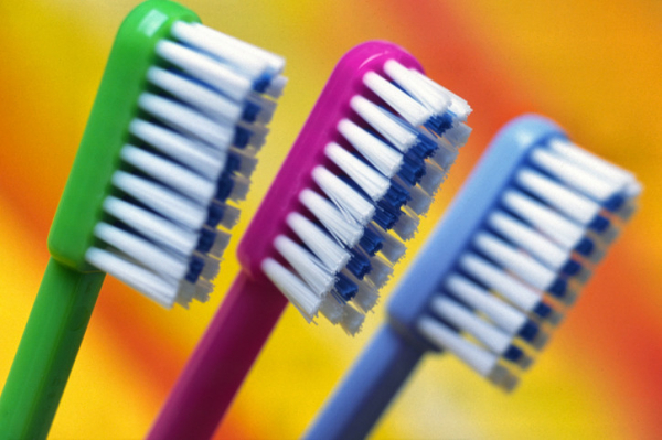
One more reason to brush your teeth?

Maybe we should add toothbrushes to the bouquet of flowers we bring to friends and family members in the hospital — and make sure to pack one if we wind up there ourselves.
New Harvard-led research published online in JAMA Internal Medicine suggests seriously ill hospitalized patients are far less likely to develop hospital-acquired pneumonia if their teeth are brushed twice daily. They also need ventilators for less time, are able to leave the intensive care unit (ICU) more quickly, and are less likely to die in the ICU than patients without a similar toothbrushing regimen.
Why would toothbrushing make any difference?
“It makes sense that toothbrushing removes the bacteria that can lead to so many bad outcomes,” says Dr. Tien Jiang, an instructor in oral health policy and epidemiology at Harvard School of Dental Medicine, who wasn’t involved in the new research. “Plaque on teeth is so sticky that rinsing alone can’t effectively dislodge the bacteria. Only toothbrushing can.”
Pneumonia consistently falls among the leading infections patients develop while hospitalized. According to the Agency for Healthcare Research and Quality, each year more than 633,000 Americans who go to the hospital for other health issues wind up getting pneumonia. Air sacs (alveoli) in one or both lungs fill with fluid or pus, causing coughing, fever, chills, and trouble breathing. Nearly 8% of those who develop hospital-acquired pneumonia die from it.
How was the study done?
The researchers reviewed 15 randomized trials encompassing nearly 2,800 patients. All of the studies compared outcomes among seriously ill hospitalized patients who had daily toothbrushing to those who did not.
- 14 of the studies were conducted in ICUs
- 13 involved patients who needed to be on a ventilator
- 11 used an antiseptic rinse called chlorhexidine gluconate for all patients: those who underwent toothbrushing and those who didn’t.
What were the findings?
The findings were compelling and should spur efforts to standardize twice-daily toothbrushing for all hospitalized patients, Dr. Jiang says.
Study participants who were randomly assigned to receive twice-daily toothbrushing were 33% less likely to develop hospital-acquired pneumonia. Those effects were magnified for people on ventilators, who needed this invasive breathing assistance for less time if their teeth were brushed.
Overall, study participants were 19% less likely to die in the ICU — and able to graduate from intensive care faster — with the twice-daily oral regimen.
How long patients stayed in the hospital or whether they were treated with antibiotics while there didn’t seem to influence pneumonia rates. Also, toothbrushing three or more times daily didn’t translate into additional benefits over brushing twice a day.
What were the study’s strengths and limitations?
One major strength was compiling years of smaller studies into one larger analysis — something particularly unusual in dentistry, Dr. Jiang says. “From a dental point of view, having 15 randomized controlled trials is huge. It’s very hard to amass that big of a population in dentistry at this high a level of evidence,” she says.
But toothbrushing techniques may have varied among hospitals participating in the research. And while the study was randomized, it couldn’t be blinded — a tactic that would reduce the chance of skewed results. Because there was no way to conceal toothbrushing regimens, clinicians involved in the study likely knew their efforts were being tracked, which may have changed their behavior.
“Perhaps they were more vigilant because of it,” Dr. Jiang says.
How exactly can toothbrushing prevent hospital-acquired pneumonia?
It’s not complicated. Pneumonia in hospitalized patients often stems from breathing germs into the mouth — germs which number more than 700 different species, including bacteria, fungi, viruses and other microbes.
This prospect looms larger for ventilated patients, since the breathing tube inserted into the throat can carry bacteria farther down the airway. “Ventilated patients lose the normal way of removing some of this bacteria,” Dr. Jiang says. “Without that ventilator, we can sweep it out of our upper airways.”
How much does toothbrushing matter if you’re not hospitalized?
In case you think the study findings only pertain to people in the hospital, think again. Rather, this drives home how vital it is for everyone to take care of their teeth and gums.
About 300 diseases and conditions are linked in some way to oral health. Poor oral health triggers some health problems and worsens others. People with gum disease and tooth loss, for example, have higher rates of heart attacks. And those with uncontrolled gum disease typically have more difficulty controlling blood sugar levels.
About the Author

Maureen Salamon, Executive Editor, Harvard Women's Health Watch
Maureen Salamon is executive editor of Harvard Women’s Health Watch. She began her career as a newspaper reporter and later covered health and medicine for a wide variety of websites, magazines, and hospitals. Her work has … See Full Bio View all posts by Maureen Salamon
About the Reviewer
Howard E. LeWine, MD, Chief Medical Editor, Harvard Health Publishing
Dr. Howard LeWine is a practicing internist at Brigham and Women’s Hospital in Boston, Chief Medical Editor at Harvard Health Publishing, and editor in chief of Harvard Men’s Health Watch. See Full Bio View all posts by Howard E. LeWine, MD

Stepping up activity if winter slowed you down

If you've been cocooning due to winter’s cold, who can blame you? But a lack of activity isn't good for body or mind during any season. And whether you're deep in the grip of winter or fortunate to be basking in signs of spring, today is a good day to start exercising. If you’re not sure where to start — or why you should — we’ve shared tips and answers below.
Moving more: What’s in it for all of us?
We’re all supposed to strengthen our muscles at least twice a week and get a total at least 150 minutes of weekly aerobic activity (the kind that gets your heart and lungs working). But fewer than 18% of U.S. adults meet those weekly recommendations, according to the CDC.
How can choosing to become more active help? A brighter mood is one benefit: physical activity helps ease depression and anxiety, for example. And being sufficiently active — whether in short or longer chunks of time — also lowers your risk for health problems like
- heart disease
- stroke
- diabetes
- cancer
- brain shrinkage
- muscle loss
- weight gain
- poor posture
- poor balance
- back pain
- and even premature death.
What are your exercise obstacles?
Even when we understand these benefits, a range of obstacles may keep us on the couch.
Don’t like the cold? Have trouble standing, walking, or moving around easily? Just don’t like exercise? Don’t let obstacles like these stop you anymore. Try some workarounds.
- If it’s cold outside: It’s generally safe to exercise when the mercury is above 32° F and the ground is dry. The right gear for cold doesn’t need to be fancy. A warm jacket, a hat, gloves, heavy socks, and nonslip shoes are a great start. Layers of athletic clothing that wick away moisture while keeping you warm can help, too. Consider going for a brisk walk or hike, taking part in an orienteering event, or working out with battle ropes ($25 and up) that you attach to a tree.
- If you have mobility issues: Most workouts can be modified. For example, it might be easier to do an aerobics or weights workout in a pool, where buoyancy makes it easier to move and there’s little fear of falling. Or try a seated workout at home, such as chair yoga, tai chi, Pilates, or strength training. You’ll find an endless array of free seated workout videos on YouTube, but look for those created by a reliable source such as Silver Sneakers, or a physical therapist, certified personal trainer, or certified exercise instructor. Another option is an adaptive sports program in your community, such as adaptive basketball.
- If you can’t stand formal exercise: Skip a structured workout and just be more active throughout the day. Do some vigorous housework (like scrubbing a bathtub or vacuuming) or yard work, climb stairs, jog to the mailbox, jog from the parking lot to the grocery store, or do any activity that gets your heart and lungs working. Track your activity minutes with a smartphone (most devices come with built-in fitness apps) or wearable fitness tracker ($20 and up).
- If you’re stuck indoors: The pandemic showed us there are lots of indoor exercise options. If you’re looking for free options, do a body-weight workout, with exercises like planks and squats; follow a free exercise video online; practice yoga or tai chi; turn on music and dance; stretch; or do a resistance band workout. Or if it’s in the budget, get a treadmill, take an online exercise class, or work online with a personal trainer. The American Council on Exercise has a tool on its website to locate certified trainers in your area.
Is it hard to find time to exercise?
The good news is that any amount of physical activity is great for health. For example, a 2022 study found that racking up 15 to 20 minutes of weekly vigorous exercise (less than three minutes per day) was tied to lower risks of heart disease, cancer, and early death.
"We don't quite understand how it works, but we do know the body's metabolic machinery that imparts health benefits can be turned on by short bouts of movement spread across days or weeks," says Dr. Aaron Baggish, founder of Harvard-affiliated Massachusetts General Hospital's Cardiovascular Performance Program and an associate professor of medicine at Harvard Medical School.
And the more you exercise, Dr. Baggish says, the more benefits you accrue, such as better mood, better balance, and reduced risks of diabetes, high blood pressure, high cholesterol, and cognitive decline.
What’s the next step to take?
For most people, increasing activity is doable. If you have a heart condition, poor balance, muscle weakness, or you’re easily winded, talk to your doctor or get an evaluation from a physical therapist.
And no matter which activity you select, ease into it. When you’ve been inactive for a while, your muscles are vulnerable to injury if you do too much too soon.
“Your muscles may be sore initially if they are being asked to do more,” says Dr. Sarah Eby, a sports medicine specialist at Harvard-affiliated Spaulding Rehabilitation Hospital. “That’s normal. Just be sure to start low, and slowly increase your duration and intensity over time. Pick activities you enjoy and set small, measurable, and attainable goals, even if it’s as simple as walking five minutes every day this week.”
Remember: the aim is simply exercising more than you have been. And the more you move, the better.
About the Author

Heidi Godman, Executive Editor, Harvard Health Letter
Heidi Godman is the executive editor of the Harvard Health Letter. Before coming to the Health Letter, she was an award-winning television news anchor and medical reporter for 25 years. Heidi was named a journalism fellow … See Full Bio View all posts by Heidi Godman
About the Reviewer
Howard E. LeWine, MD, Chief Medical Editor, Harvard Health Publishing
Dr. Howard LeWine is a practicing internist at Brigham and Women’s Hospital in Boston, Chief Medical Editor at Harvard Health Publishing, and editor in chief of Harvard Men’s Health Watch. See Full Bio View all posts by Howard E. LeWine, MD